Evaluating an Investigational MRI Contrast Agent for CNS & Body Imaging
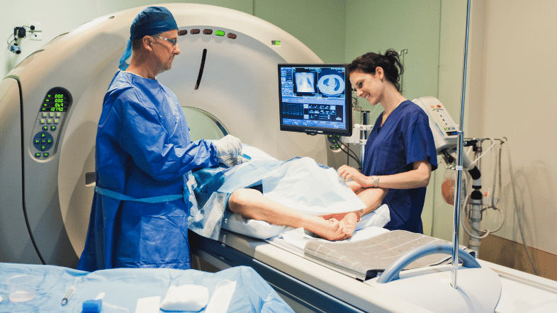
MRI is central to CNS development because it provides sensitive, non-invasive lesion visualisation and quantitative biomarkers that inform both early go/no-go decisions and late-phase efficacy. In this anonymised case, a biotech sponsor partnered with Aixial to evaluate an investigational MRI contrast agent (‘investigational agent’) against a standard reference agent (‘standard agent’) in adults; and to assess added diagnostic value versus unenhanced MRI in children. Aixial led the analysis strategy, blinded reader framework, PMDA compliant CDISC standardisation, producing a submission-grade evidence package for global health-authority pathways – without disclosing proprietary product identifiers or results.
Date: 13 January 2026 | Ref: ART015
Neurological pain disorders—such as neuropathic pain, migraine, and fibromyalgia—remain among the most difficult indications in central nervous system (CNS) drug development. Pain trials are failing not because the science is weak, but because the model of clinical development for neurological pain has not evolved in over two decades.
How Aixial’s imaging and biostatistics teams delivered regulator-ready analyses across adult and paediatric cohorts
Compliance note: This case study intentionally does not name the investigational product or comparator and does not report sponsor results. Content focuses on study design, methods, and CRO value delivery.
Study at a Glance
Populations:
- Adults: 200 participants (including 80 with CNS indications)
- Paediatrics: 40 participants (including 19 with CNS indications)
Design
- Adults: Randomised, double-blind, cross-over
- Paediatrics: Open-label, single-arm
Primary Objectives:
- Adults: Demonstrate non-inferiority of the investigational agent vs the standard agent for lesion visualisation across CNS and other body regions
- Paediatrics: Evaluate added diagnostic value of the investigational agent vs unenhanced MRI
Blinded Evaluation: 3 independent radiologists per body region, pooled as Reader A/B/C, to mitigate single-reader bias
Decision Framework: 10% non-inferiority margin (0.35 on an expected mean of 3.5); non-inferiority declared if all 3 co-primary criteria are met simultaneously for ≥ 2 of 3 readers
Sensitivity & Subgroups: Worst-score imputation for non-matching lesions; alternate reader poolings; CNS vs non-CNS subgroup analyses
Reliability: Intra- and inter-reader ICC to quantify repeatability and agreement
Study Design Overview — Investigational MRI Contrast Agent (CNS & Body)
| Section | Adults N=200 (80 CNS) | Paediatrics N=40 (19 CNS) |
| Design | Randomised, double-blind, cross-over Open-label, single-arm Primary objective Non-inferiority vs. standard agent for lesion visualisation (CNS & other regions) | Added diagnostic value vs. unenhanced MRI for lesion visualisation |
| Lesion visualisation assessment | 3 co-primary criteria assessed on a 4-point scale; patient-level mean across up to 10 largest & most enhancing lesions | 3 criteria assessed on a 4-point scale; same lesion-level rules where applicable |
| Blinded evaluation | 3 independent radiologists per body region; pooled as Readers A/B/C | 3 independent radiologists per body region; pooled as Readers A/B/C |
| Reader pooling robustness | Secondary analyses with alternate random combinations of readers | |
| Non-inferiority framework | Margin 10% (0.35 on expected mean 3.5); NI if all 3 co-primary criteria met for ≥2/3 readers | Not applicable (no NI hypothesis; focuses on added value vs. unenhanced) |
| Sensitivity analyses | Worst-score imputation for lesions not seen with investigational agent; include matching & non-matching lesions | Worst-score imputation approach mirrored where relevant to paediatric objective |
| Subgroups | CNS (brain & spine) vs. other body regions | CNS (brain & spine) vs. other body regions |
| Reliability | Intra- and inter-reader variability assessed via ICC (target: good-to-excellent) | Inter-reader variability assessed via ICC (target: good-to-excellent) |
Why This Matters to Biotech Sponsors
Lesion visualisation is often the fulcrum for value in CNS imaging trials. Sponsors need confidence that endpoints are clinically justified (margin, scales, lesion multiplicity), reader variability is engineered and measured, analyses are regulator-ready (CDISC, traceable derivations, reproducible TFLs), and the narrative scales into pooled safety/efficacy summaries for streamlined review. Aixial addressed these needs with a design-forward and submission-back approach – planning endpoints and reader strategy up front, while building data flows that map cleanly into global submissions later.
What Aixial Delivered
1) Analysis Strategy & Reader Science
- Lesion-level rules (up to the 10 largest and most enhancing lesions) pre-specified to reflect clinical reality and control multiplicity.
- Blinded, independent readers per body region with pooling to Reader A/B/C; three secondary analyses with alternate reader combinations to demonstrate invariance of conclusions.
- Reliability quantified through intra/inter-reader ICC, with reader training and calibration documented.
2) Non-Inferiority Framework that Stands Up to Scrutiny
- Clear clinical rationale for a 10% margin (expressed on a 4-point scale as 0.35) tied to the practical meaning of ‘no clinically important difference’ in lesion visualisation.
- Decision rule requiring concordance across three co-primary criteria for ≥ 2 of 3 readers, increasing robustness to reader-specific effects.
3) Submission-Grade Standards & Traceability
- PMDA compliant CDISC SDTM & ADaM datasets verified with Pinnacle 21; eCRT packages; fully documented Table Figures and Listings (TFLs).
- Pooled analyses: Integrated summaries for safety and efficacy assembled from multiple studies to support a cohesive submission narrative.
- Audit-ready lineage: Every endpoint and table traceable back to specifications, derivations, and source data.
4) Pediatric-Specific Considerations
- Ethical guardrails (consent/assent), dose selection rationale for contrast administration, and a design suited to clinical feasibility (single-arm vs unenhanced baseline).
Best Practices We Recommend (and Implemented Here)
- Pre-specify sensitivity pathways (e.g., worst-score imputation for lesions not detected by one agent) before any unblinding.
- Engineer reliability: reader calibration sessions, qualification criteria, and ICC thresholds (e.g., good ≥ 0.75).
- Power cross-over designs properly: include within-subject correlation, sequence balance, and carryover mitigation.
- Design for submissions from Day 1: CDISC mapping plans, reusable TFL shells, and pooled-summary strategy to cut rework and shorten review cycles.
- Make the endpoint interpretable: tie the margin and scales back to clinical decision-making, not just statistics.
Engagement Options for Biotech Teams
- End-to-end package: Protocol consult → SAP/Readers → CDISC → ISS/ISE → Submission support.
- Modular resourcing: Plug-in biostatistics, imaging oversight, or data standards as standalone services.
- Quality guardrails & KPIs: Pinnacle 21 issue thresholds, read turnaround SLAs, QC coverage %, and reproducibility checks.
Let’s design a reader-robust, regulator-ready imaging plan for your next CNS study.
TO GO FURTHER




How can we support your next project?
Whether you’re looking for a protocol review or a proposal,
simply reach out to us by filling our request for proposal.